
If you’ve been dealing with stubborn back or buttock pain and someone has mentioned ankylosing spondylitis (AS),
you’re probably wondering whether an MRI can finally “catch it in the act.” The short version: yes,
ankylosing spondylitis (and its close cousin, axial spondyloarthritis) can show up on MRIsometimes before it shows up on X-ray.
The longer version is more nuanced (because of course it isyour spine didn’t get the memo that you like simple answers).
This article breaks down what doctors look for, what “positive” MRI findings mean, why MRI can still miss things,
and how to read an MRI report without spiraling into a late-night search frenzy. (Okay, maybe just a smaller frenzy.)
First, a quick translation: AS, axSpA, and why names matter
Ankylosing spondylitis is part of a family of inflammatory conditions called axial spondyloarthritis (axSpA).
The “axial” part means it mainly targets the spine and the sacroiliac (SI) jointsthe joints where your spine meets your pelvis.
You’ll often hear two buckets:
- Radiographic axSpA (classic ankylosing spondylitis): structural damage is visible on X-ray, especially in the SI joints.
-
Non-radiographic axSpA: symptoms and inflammation may be present, but X-rays don’t yet show “definitive” SI-joint damage.
MRI is frequently used here because it can detect inflammation earlier.
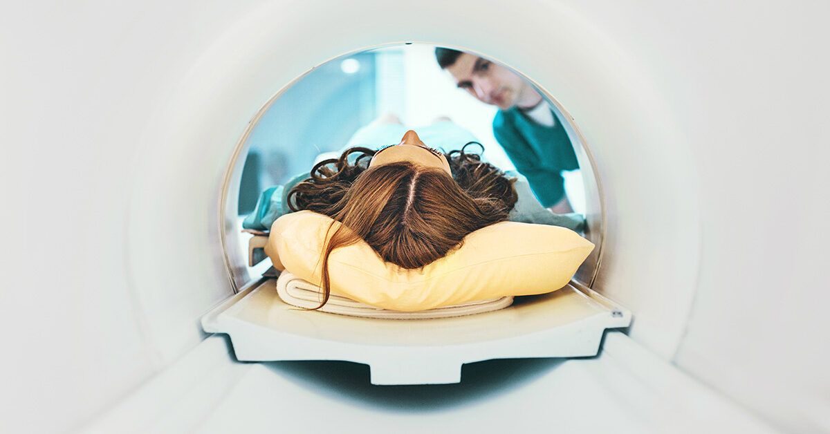
So… does ankylosing spondylitis show up on MRI?
Often, yes. MRI can show signs of inflammation in the SI joints and sometimes the spineparticularly in earlier disease,
when X-rays may look normal. MRI is good at spotting active inflammation (think swelling and inflammatory changes in bone and soft tissue),
as well as some structural changes (damage or remodeling) depending on the sequences used.
But here’s the catch: MRI is not a perfect “yes/no” test for AS. You can have axial spondyloarthritis with a normal MRI,
and you can have MRI findings that look suspicious but are caused by something else.
Why MRI can be a big deal (especially early on)
Traditional X-rays are great at showing bone changeserosions, sclerosis, joint space changes, and eventually fusion.
The problem is that those changes can take years to become obvious. MRI, on the other hand, can detect inflammation sooner,
potentially shortening the long “diagnosis limbo” many people experience.
In practical terms, MRI can help when:
- You have chronic back pain with features that sound inflammatory (more on that soon), but X-rays are normal.
- Your clinician suspects non-radiographic axial spondyloarthritis and wants evidence of active inflammation.
- There’s uncertainty and the goal is to support (or challenge) the diagnosis alongside symptoms and labs.
- You need a clearer picture of what’s happening in the SI joints or spine to guide treatment decisions.
What doctors look for on MRI
Most MRI discussions about ankylosing spondylitis revolve around the SI joints. That’s because inflammation therecalled
sacroiliitisis a hallmark feature in axial spondyloarthritis.
1) Active inflammatory changes (the “right now” clues)
The most talked-about MRI finding is bone marrow edema (also described as osteitis) near the SI joints.
On fluid-sensitive MRI sequences (often STIR or fat-suppressed T2), this can appear as bright areas in the bone just next to the joint.
Other “active” findings may include:
- Synovitis (inflammation of the joint lining)
- Capsulitis (inflammation of the joint capsule)
- Enthesitis (inflammation where tendons/ligaments attach to bone)
- Soft tissue inflammation around the joint
2) Structural changes (the “history book” clues)
MRI can also show longer-term changes, especially on T1-weighted images. These may include:
- Erosions (small areas where bone has been worn away)
- Fat metaplasia (fatty replacement in bone marrow after inflammation)
- Sclerosis (hardening/thickening of boneoften seen better on X-ray or CT but can be suggested on MRI)
- Ankylosis (fusionmore advanced disease)
Structural findings can strengthen the case when inflammation is subtle, but interpretation depends on the overall clinical picture.
What about the spine?
Yes, MRI can show inflammatory lesions in the spine toosometimes called “corner lesions” or inflammatory changes at vertebral edges.
However, SI-joint MRI is often prioritized in suspected early disease because sacroiliitis is such a key diagnostic feature.
Spine MRI may be considered when symptoms strongly suggest axial spondyloarthritis but SI-joint imaging is inconclusive, or when
clinicians are evaluating complications or alternate causes of pain.
When MRI might NOT show ankylosing spondylitis (and why that’s not the end)
A normal MRI can be frustratingespecially if your pain is real (it is) and persistent (also yes).
Here are common reasons MRI may come back “unremarkable” even when axial spondyloarthritis is still on the table:
The inflammation can be patchy or quiet that day
Inflammatory disease doesn’t always flare 24/7. If the scan happens when inflammation is low, the classic edema patterns may not appear.
Timing and treatment can change the picture
Anti-inflammatory medications and biologic therapies can reduce MRI-visible inflammation. That’s good for your body,
but it can make diagnostic imaging less dramatic.
The wrong area (or the wrong protocol) was scanned
A generic “lumbar spine MRI” done for sciatica may not adequately image the SI joints. When the clinical question is axial spondyloarthritis,
the request often needs to explicitly mention SI-joint MRI and use sequences sensitive to inflammation.
Radiology interpretation is specialized
Subtle sacroiliitis can be difficult to read. Radiologists and rheumatologists familiar with spondyloarthritis imaging
are better positioned to interpret borderline findings.
Can MRI be “positive” for the wrong reasons?
Yesand this is where things get interesting (and mildly annoying).
Some MRI findings that resemble inflammatory sacroiliitis can occur in other situations, including:
- Mechanical stress (certain sports, heavy training, repetitive strain)
- Postpartum changes (pregnancy and childbirth can stress SI joints and may cause temporary inflammatory-looking changes)
- Degenerative arthritis (wear-and-tear patterns can overlap in spots)
- Infection (less common but important to rule out when symptoms and labs suggest it)
- Stress injuries or fractures
This is why good clinicians don’t diagnose AS from an MRI image alone. They combine MRI findings with symptoms,
exam findings, labs, and sometimes repeat imaging over time.
MRI vs X-ray vs CT: which test shows what?
X-ray
X-rays show structural damagebut often later in the disease course. They’re commonly used to look for
“definitive” sacroiliitis in established ankylosing spondylitis and to track progression over time.
MRI
MRI excels at showing inflammation and can detect earlier disease (especially non-radiographic axSpA),
with no radiation exposure. It can also show some structural changes, but interpretation depends on technique and expertise.
CT
CT is excellent for detailed bone anatomy and can reveal erosions or bony changes more clearly than X-ray in some cases.
However, it uses radiation and is not typically the first-line test for early inflammatory detection.
What an MRI report might say (in human terms)
Radiology reports can sound like they were written by a committee of robots arguing over commas. Here are some common phrases
and what they may mean in plain English:
-
“Bone marrow edema involving the iliac side of the SI joint”:
inflammation-type signal in the pelvic bone near the joint; may support sacroiliitis depending on pattern and context. -
“Findings suggestive of active sacroiliitis”:
the radiologist sees a pattern that fits inflammatory disease; clinicians will correlate with symptoms and labs. -
“No evidence of sacroiliitis”:
no obvious inflammatory pattern was seendoes not always rule out axSpA, especially if clinical suspicion remains high. -
“Erosions and/or fatty metaplasia”:
evidence of prior or chronic inflammatory changes; can add weight to the diagnosis. -
“Degenerative changes”:
wear-and-tear findings; may explain pain, or may coexist with inflammatory disease.
How to increase the odds your MRI actually answers the right question
You don’t need to micromanage your healthcare teambut being informed helps. Consider these practical tips:
- Make sure the SI joints are included. If the question is axial spondyloarthritis, a dedicated SI-joint MRI is often needed.
- Ask whether inflammation-sensitive sequences are used. Many protocols include STIR or fat-suppressed T2 plus T1 images.
-
Share relevant symptoms. Night pain, morning stiffness, improvement with exercise, alternating buttock pain,
uveitis history, psoriasis, or inflammatory bowel disease can all matter. - Consider rheumatology involvement early. Rheumatologists specialize in interpreting the whole puzzle, not just one test.
-
Don’t panic if the MRI is normal. A normal scan can be useful information, and next steps may include follow-up,
different imaging, or re-evaluating alternate diagnoses.
How clinicians connect MRI findings to a real diagnosis
In many clinics, MRI evidence of sacroiliitis can support classification criteria used in research and help guide clinical thinking
but doctors still make a diagnosis based on the full picture:
- Symptoms: inflammatory back pain pattern (often starting before age 45, lasting >3 months, improving with activity)
- Physical exam: tenderness, range-of-motion limits, chest expansion changes, functional limitations
- Blood tests: HLA-B27 (not definitive), CRP/ESR (may be normal even in active disease)
- Imaging: X-ray and/or MRI of SI joints (and sometimes spine)
- Extra features: uveitis, psoriasis, inflammatory bowel disease, enthesitis, family history
In other words: MRI is a powerful piece of evidence, not the whole courtroom drama.
FAQ: common questions people ask after Googling “AS MRI” at 1 a.m.
Does ankylosing spondylitis always show on MRI?
No. Some people with early axial spondyloarthritis may have a normal MRI, especially if inflammation is low at the time of imaging,
if the SI joints weren’t properly evaluated, or if findings are subtle.
Can MRI detect AS earlier than X-ray?
Often yesespecially for inflammation. X-rays are better for later structural changes; MRI can identify earlier inflammatory changes in the SI joints.
Do you need contrast (gadolinium) for AS MRI?
Many SI-joint MRI protocols can detect key inflammatory findings without contrast. In certain scenarios, contrast may help clarify synovitis or active inflammation,
but it’s not always required. Your clinician and radiology team decide based on the question being asked.
What if the MRI shows sacroiliitisdoes that automatically mean AS?
Not automatically. Sacroiliitis can have different causes. The MRI pattern, your symptoms, exam, and lab results help determine whether it fits axial spondyloarthritis.
Bottom line: what to do with your MRI result
If your MRI shows inflammatory sacroiliitis, it can strongly support a diagnosis of axial spondyloarthritisespecially when symptoms match.
If your MRI is normal, it doesn’t necessarily mean “nothing is wrong.” It may mean the inflammation isn’t visible right now, the wrong area was imaged,
or another condition is responsible for your symptoms.
The best next step is usually the least dramatic one: review results with a clinician (often a rheumatologist),
talk through your symptom pattern and medical history, and decide whether you need additional testing, follow-up, or a different plan.
And yes, you’re allowed to ask for the report explained in plain English. It’s your body; you get the user manual.
Real-World Experiences: What It Can Feel Like When AS Meets MRI (Extra ~)
Let’s talk about the part that doesn’t show up neatly in a radiology report: the experience of getting from “my back hurts”
to “here’s what’s actually going on.”
Many people describe the early phase as a slow-burn mystery. It’s not the dramatic “I lifted a couch wrong” back pain.
It’s the kind that creeps in, hangs out, andrudelyoften feels worse after resting. Folks commonly report waking up stiff,
needing a hot shower to feel human, and noticing that movement helps more than lying down. That’s a weird clue, by the way:
most back pain loves rest; inflammatory back pain tends to get cranky when you’re still.
Then comes the healthcare scavenger hunt. A typical path might include physical therapy, posture lectures, new pillows,
and at least one person (well-meaning, but incorrect) telling you to “just strengthen your core.” Some people get told it’s a muscle strain,
a disc issue, or stress. Othersespecially those whose symptoms don’t match the classic stereotypedescribe bouncing between explanations
before anyone mentions inflammatory arthritis. That delay can feel validating (“I’m not imagining it”) and frustrating (“Why did this take so long?”)
at the same time.
When MRI enters the story, expectations can get sky-high. People often hope it will deliver a crystal-clear verdict:
Yes, it’s AS or No, it’s not. In reality, MRI results can feel like reading a horoscope written by a scientist:
“Findings may be suggestive… correlate clinically.” Translation: “We see something that could fit, but your doctor needs the full context.”
Some patients describe relief when the report notes sacroiliitisfinally, proof. Others feel crushed when the report says everything looks normal
despite daily pain.
A surprisingly common experience is learning that a normal MRI doesn’t mean a normal life. People may still have inflammatory symptoms,
flares that come and go, and fatigue that doesn’t care about your calendar. In those cases, clinicians might monitor symptoms over time,
repeat imaging later, or look closely at other evidence (like HLA-B27 status, inflammatory markers, and extra symptoms such as eye inflammation).
For many, the turning point is seeing a rheumatologist who treats the whole pattern rather than a single test result.
Practical tip from the lived-experience department: bring notes to appointments. People who document symptom timing (morning stiffness duration,
night pain, response to NSAIDs, flare triggers) often feel more confident advocating for themselvesand clinicians get clearer data.
Also, don’t be shy about asking, “Was the MRI focused on the SI joints?” Plenty of patients discover they had a lumbar spine MRI that
didn’t really answer the spondyloarthritis question.
Finally, there’s the emotional side. Whether your MRI is positive, negative, or “meh,” it’s normal to feel a mix of hope, anger, relief,
and exhaustion. The goal isn’t a perfect scanit’s a plan that reduces pain, protects mobility, and gets you back to living your life
with fewer negotiations between you and your spine. (May your spine eventually agree to reasonable terms.)