
When your doctor says, “We’d like to order a bone scan,” it can sound a little dramatic, like your skeleton just got promoted to lead actor. In reality, a bone scan is a common imaging test that helps doctors spot areas where your bones are unusually active. That matters because bones are not static sticks holding you upright like coat hangers. They are living tissue, constantly breaking down, rebuilding, repairing, and occasionally complaining.
A bone scan does not simply take a photograph of your bones the way a regular X-ray does. Instead, it shows bone activity. In other words, it can reveal where bone is healing, inflamed, irritated, infected, or affected by cancer. That difference is the whole plot twist. If an X-ray is a still photo, a bone scan is more like a heat map of what your skeleton is doing behind the scenes.
This article explains what a bone scan shows, what the results can mean, how the test works, and how it differs from a bone density scan. If you have an upcoming appointment, think of this as the calm, clear friend who sits next to you in the waiting room and tells you what’s actually going on.
What is a bone scan?
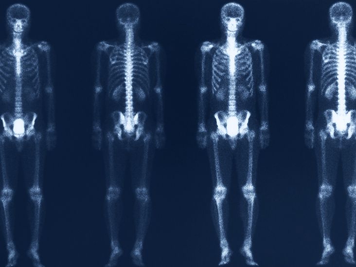
A bone scan, also called bone scintigraphy, is a type of nuclear medicine imaging test. A small amount of radioactive tracer is injected into a vein, usually in your arm or hand. The tracer travels through your bloodstream and collects in your bones. A special camera, often called a gamma camera, detects the tracer and creates images that show how actively different parts of your skeleton are functioning.
The key idea is simple: areas of bone that are changing quickly tend to absorb more tracer. These show up as hot spots. Areas that absorb less tracer may appear as cold spots. Neither one is a diagnosis by itself. They are clues. Your doctor reads those clues alongside your symptoms, exam findings, blood work, and other imaging tests such as X-rays, CT scans, MRI, or sometimes PET scans.
What does a bone scan show?
A bone scan is especially good at showing abnormal bone metabolism or increased bone turnover. That means it can help reveal places where bone is repairing itself or reacting to disease. Instead of only asking, “Does this bone look different?” the scan asks, “Is this bone behaving differently?”
Here’s what a bone scan may show:
- Fractures, especially tiny stress fractures or breaks that are hard to see on a standard X-ray early on
- Bone infection such as osteomyelitis
- Arthritis and joint changes, including areas where wear-and-tear or inflammation is especially active
- Cancer that has spread to bone, also called bone metastases
- Primary bone tumors or suspicious bone lesions that need more evaluation
- Unexplained bone pain when other tests have not given a clear answer
- Problems around prosthetic joints, such as loosening or abnormal bone activity after joint replacement
- Reduced blood supply to bone in some situations, including conditions such as osteonecrosis
In cancer care, a whole-body bone scan is often used because it can look at the entire skeleton in one session. That makes it useful when doctors need to know whether cancer has spread beyond its original site. A localized scan, on the other hand, may focus on one area, such as the knee, hip, spine, or foot.
What do hot spots and cold spots mean?
Hot spots
Hot spots are areas where the tracer collects more heavily. This usually means the bone in that area is more metabolically active than expected. That may happen when bone is healing after a fracture, reacting to arthritis, responding to infection, or affected by tumor activity. Hot spots are common, but they are not specific. One bright area on a scan does not automatically mean cancer. Bones, like people on deadline, can get active for many reasons.
Cold spots
Cold spots are areas that take up less tracer than surrounding bone. These may signal reduced blood flow, certain types of lesions, or another process that lowers normal tracer uptake. They are less common than hot spots, but they matter. A radiologist will consider the pattern, location, and your medical history before deciding what it may mean.
Why results are not read in isolation
A bone scan is sensitive, which means it can detect abnormal activity well. But it can also be nonspecific, which means different conditions can create similar-looking scan patterns. For example, a healing fracture, arthritis flare, and metastatic lesion may all produce increased uptake. That is why doctors often pair a bone scan with X-rays, MRI, CT, SPECT/CT, lab tests, or follow-up imaging.
Conditions a bone scan can help detect
1. Stress fractures and hidden breaks
If you have persistent foot, shin, hip, or back pain and the X-ray looks normal, a bone scan may help show whether the bone is quietly trying to repair a small crack. This can be especially helpful in athletes, runners, military recruits, and anyone whose hobbies accidentally qualify as full-contact cardio.
2. Bone metastases
One of the best-known uses of a bone scan is checking whether cancers, especially cancers such as prostate, breast, or lung cancer, may have spread to bone. Because a whole-body scan surveys the skeleton, it can reveal multiple active sites that would be easy to miss if only one painful area were imaged.
3. Bone infection
A bone scan may help identify osteomyelitis when infection is suspected, particularly if symptoms, lab results, or other imaging point in that direction. Sometimes additional specialized scans or MRI are needed for better detail, but the bone scan can be part of the diagnostic puzzle.
4. Arthritis and degenerative change
Arthritic joints often show increased tracer uptake because the bone near the joint is remodeling more actively. A scan can sometimes help pinpoint which joints are driving symptoms, especially when someone has pain in multiple places and a less obvious source.
5. Problems after joint replacement
Doctors may use a bone scan when they suspect abnormal bone activity around a prosthetic hip or knee, such as loosening, stress reaction, or complications that need a closer look.
What a bone scan does not show well
A bone scan is useful, but it is not magic and it is definitely not a psychic reading for your skeleton.
- It does not tell the exact cause of every abnormal spot by itself.
- It does not show soft tissues as clearly as MRI.
- It does not replace CT when detailed anatomy is needed.
- It does not measure bone density or diagnose osteoporosis the way a DEXA scan does.
That last point causes a lot of confusion. A bone scan and a bone density scan are completely different tests. They share the word “bone,” which is not exactly helpful, but they answer different questions.
Bone scan vs. bone density scan: not the same thing
A bone scan looks for abnormal activity in the skeleton. It can help detect fractures, infection, arthritis, tumors, and cancer spread.
A bone density scan, often called DEXA or DXA, measures how much mineral is packed into your bones. It is primarily used to screen for osteoporosis and estimate fracture risk. It does not show hot spots. It does not map active disease the way a nuclear bone scan does.
So if someone says, “I had a bone scan and they checked me for osteoporosis,” they may actually mean they had a DEXA scan. Medical terminology loves a good mix-up.
How the test is performed
Step 1: The tracer injection
You receive a small amount of radiotracer through an IV injection. This part is quick and usually feels like a routine blood draw or IV placement.
Step 2: The waiting period
After the injection, there is usually a wait of about one to four hours while the tracer circulates and settles into your bones. You may be asked to drink water during this time to help clear excess tracer from your body. Depending on the reason for the test, some early images may also be taken right after the injection.
Step 3: The scan itself
When imaging starts, you lie on a table while the camera moves around or above your body. The scan often takes roughly 30 to 60 minutes, though timing varies. You need to stay as still as possible so the images do not blur. If holding one position is difficult because of pain, tell the staff before the scan. That is a practical detail, not a personality flaw.
Step 4: Additional imaging if needed
Sometimes the exam includes SPECT or SPECT/CT. These techniques create more detailed, three-dimensional images and can help localize exactly where abnormal uptake is happening. That can be especially useful in complex areas like the spine, pelvis, or joints.
How to prepare for a bone scan
Preparation is usually simple, but you should follow your imaging center’s specific instructions. In general:
- Tell your care team if you are pregnant, might be pregnant, or are breastfeeding.
- Wear comfortable clothes and expect to remove jewelry or metal items.
- Tell the staff about medications, vitamins, allergies, and recent imaging tests.
- Ask ahead if you have trouble lying flat, significant pain, or claustrophobia.
Most people do not need major diet changes or intense preparation. This is not one of those tests where you have to memorize a four-page instruction sheet and question your life choices.
Are bone scans safe?
For most people, yes. Bone scans use a small amount of radiation. Serious side effects are uncommon. The radiotracer injection may briefly cause mild discomfort, and allergic reactions are rare and usually mild. Many patients are advised to drink extra fluids afterward so the tracer leaves the body more quickly.
That said, bone scans are not automatically appropriate during pregnancy, and breastfeeding may require special guidance depending on the tracer and facility protocol. If either applies to you, bring it up before the test, not after you are already halfway into the gown and wondering why hospital socks have such strong opinions.
What do the results mean?
A normal bone scan usually shows tracer distributed in a balanced way throughout the skeleton, without unusual hot or cold spots.
An abnormal bone scan means there are areas of higher or lower uptake than expected. The meaning depends on the pattern:
- Single hot spot: may suggest a localized issue such as a fracture, arthritis, or infection
- Multiple hot spots: may suggest widespread bone involvement, including metastatic disease in the right clinical context
- Cold spot: may indicate an area that needs more investigation because uptake is reduced
- Symmetric uptake near joints: can be consistent with degenerative or inflammatory joint changes
The most important thing to remember is this: a bone scan result is rarely the final word. It is often the beginning of a more precise conversation. Your doctor may recommend follow-up imaging or correlation with symptoms and lab results before making a diagnosis.
Experiences related to “Bone scan: What does it show?”
Many people worry about the wrong part of a bone scan. They imagine the machine will be the hardest part, when in reality the biggest surprise is often the waiting period between the injection and the pictures. Patients commonly say the procedure is more boring than painful. The injection is brief, the scan table is manageable, and the main challenge is simply staying still long enough to let the camera do its job.
People being scanned for unexplained pain often go in hoping for a clean, simple answer. Sometimes the scan delivers one. A runner with persistent shin pain may finally learn that the issue is a stress fracture that never showed up clearly on X-ray. A person with stubborn foot pain may discover that the “just walk it off” strategy was not the inspirational wellness advice they thought it was. In these cases, the bone scan helps validate symptoms that were real all along.
For people with a history of cancer, the emotional experience can be very different. A whole-body bone scan may feel less like routine imaging and more like a major checkpoint. Patients often describe the test as physically straightforward but emotionally heavy. The pictures are looking for whether disease has spread, and that possibility naturally raises the stakes. Even so, many patients also say that having a full-body test brings a strange kind of relief. It gives their medical team a broader view and helps guide the next step instead of leaving everyone guessing.
Another common experience comes from people with arthritis or joint replacements. They may have pain in several places at once and struggle to identify what is actually driving the problem. In that setting, the bone scan can act like a spotlight, showing which area is most metabolically active and deserves closer attention. It does not solve every mystery on its own, but it helps narrow the list of suspects.
Patients being evaluated for bone infection often report feeling frustrated before the scan because symptoms may be vague or overlap with other issues. A bone scan can help move the workup forward, especially when the source of pain, swelling, or fever is not obvious. It may not be the only test they need, but it can be the moment the investigation stops wandering in circles.
From a practical standpoint, people usually remember three things about the day of the test: bring patience, drink water if instructed, and wear something comfortable. If back pain or joint pain makes lying flat difficult, mentioning that early can make the experience much smoother. Imaging staff deal with this all the time, and small adjustments can make a big difference.
In the end, most patients come away saying the same thing: the name “bone scan” sounded scarier than the test itself. What it shows is not just whether a bone looks normal or abnormal, but whether it is actively reacting to injury or disease. That is why doctors still use it. It gives your skeleton a chance to tell its side of the story, which, honestly, it has probably been trying to do for a while.
Final takeaway
A bone scan shows how active your bones are, not just how they look. It can highlight areas of healing, inflammation, infection, arthritis, tumor activity, or cancer spread. It is especially helpful when doctors need to look at the whole skeleton or investigate symptoms that are real but not yet obvious on standard imaging.
If your doctor orders one, the smartest question is not “Should I panic?” but “What question is this test trying to answer?” Once you know that, the scan makes much more sense. It is not a random detour. It is a targeted tool that helps connect symptoms, biology, and next steps.