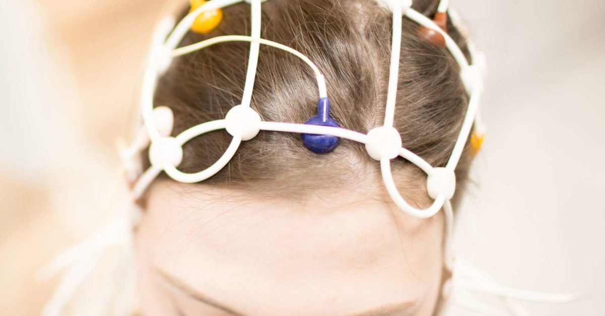
An electroencephalogram, better known as an EEG, records the electrical activity produced by the brain. The name may sound like a machine that belongs in a science-fiction laboratory, but the procedure is surprisingly straightforward: small sensors are attached to the scalp while a computer records brain-wave patterns.
Doctors most commonly order an EEG when they suspect seizures or epilepsy, although the test can also provide useful information about unexplained fainting, confusion, sleep-related events, brain injuries, infections, coma, and other neurological concerns. The test is generally safe, painless, and noninvasive. The electrodes only detect signals; they do not send electricity into the brain.
Still, an EEG is not a magical mind-reading helmet. It cannot reveal thoughts, measure intelligence, or always provide an instant diagnosis. Understanding what the test canand cannotshow makes the experience far less mysterious.
What Is an EEG Test?
Brain cells communicate through tiny electrical impulses. When groups of brain cells are active together, their combined electrical activity produces patterns called brain waves. An EEG captures these patterns through electrodes placed at carefully measured locations on the scalp.
The electrodes are connected by thin wires to an amplifier and recording system. The signals are displayed as a series of wavy lines, known as EEG tracings, on a computer screen. Each channel represents electrical activity detected from a particular region of the head. A neurologist or another physician trained in clinical neurophysiology later reviews the recording for expected rhythms, unusual slowing, spikes, sharp waves, or other potentially significant patterns.
An EEG measures brain function over time, while imaging tests such as magnetic resonance imaging, or MRI, primarily show brain structure. In simple terms, an MRI takes a detailed picture of the brain’s anatomy; an EEG listens to its electrical conversation.
Why Might a Doctor Order an EEG?
Seizure evaluation is the best-known use of electroencephalography. A doctor may recommend an EEG after an episode involving loss of awareness, unexplained staring, unusual movements, sudden confusion, muscle jerking, a blackout, or a suspected first seizure.
Diagnosing and Classifying Seizures
EEG findings may help determine whether an event was associated with abnormal electrical activity and whether the pattern appears focal, meaning it begins in one area, or generalized, meaning it involves networks on both sides of the brain. This information may help clinicians classify a seizure disorder and select appropriate treatment.
Video may be recorded at the same time so the medical team can compare physical behavior with brain activity. This is especially helpful when doctors need to distinguish epileptic seizures from fainting, sleep movements, migraine symptoms, panic episodes, movement disorders, or functional nonepileptic events.
Investigating Other Neurological Problems
An EEG may also be used when evaluating:
- Unexplained periods of confusion or altered awareness
- Fainting spells or episodes of memory loss
- Encephalitis and other conditions affecting brain function
- Head injury, stroke, or suspected brain damage
- Sleep disorders and unusual nighttime behaviors
- Critically ill patients who may be having seizures without visible shaking
- Brain activity during certain operations
- Coma or severe disorders of consciousness
In selected critical-care situations, EEG may be one component of a broader medical evaluation of brain function. It is not automatically required in every case, and it is interpreted alongside the patient’s examination, history, imaging, laboratory results, and other tests.
Types of EEG Tests
The appropriate EEG depends on how often symptoms occur, whether sleep is important, and what information the medical team needs.
Routine EEG
A routine EEG is usually performed in a clinic, hospital, or neurodiagnostic laboratory. The actual recording often lasts about 20 to 40 minutes, although the full appointment may take an hour or longer because the technologist must measure the head, prepare the scalp, apply the electrodes, perform the recording, and remove the equipment.
Sleep or Sleep-Deprived EEG
Some abnormal patterns are more likely to appear during drowsiness or sleep. A patient may therefore be asked to sleep less than usual before the appointment. Sleep deprivation can also increase the likelihood of recording certain epileptiform patterns.
Follow the laboratory’s instructions exactly. Being told to reduce sleep does not mean staying awake all night unless the care team specifically says so. A sleep-deprived person may also need someone else to provide transportation.
Ambulatory EEG
An ambulatory EEG uses a small portable recorder to monitor brain activity at home, commonly for 24 to 72 hours. The electrodes remain attached while the patient completes much of their normal routine. A diary and an event button may be used to mark symptoms, activities, medications, and sleep periods.
A longer recording can be useful when unusual electrical activity occurs too infrequently to appear during a short routine test. Patients generally cannot shower, swim, or allow the recorder to become wet while the equipment is attached.
Video EEG Monitoring
Video EEG synchronizes a brain-wave recording with video and audio. It may be conducted at home or in an epilepsy monitoring unit. Inpatient monitoring can continue for several days when clinicians need to capture a typical event, evaluate difficult-to-control seizures, adjust medication under supervision, or prepare for epilepsy surgery.
Continuous EEG
Continuous EEG is often used in intensive care units to look for ongoing or repeated seizures that may not produce obvious body movements. For example, a critically ill person may appear unusually sleepy or confused while abnormal electrical activity continues in the brain.
How to Prepare for an EEG
Your testing center’s instructions take priority because preparation varies according to the type of EEG, your age, your medications, and whether sedation is planned.
Wash Your Hair
Wash and dry your hair before the appointment, usually the night before or the morning of the test. Do not apply conditioner, oil, gel, styling cream, hairspray, or other hair products unless the laboratory says otherwise. Clean, product-free hair helps the electrodes maintain good contact with the scalp.
Patients with hair extensions, braids, weaves, wigs, scalp conditions, or adhesive allergies should contact the laboratory ahead of time. The team may provide additional instructions or modify the attachment method.
Ask About Food, Caffeine, and Sleep
Many centers advise avoiding caffeine for several hours before an EEG, particularly when sleep is part of the test. Instructions may include coffee, tea, cola, energy drinks, chocolate, and certain medications containing caffeine.
Unless instructed to fast, patients can often eat normally. A balanced meal may help prevent low blood sugar, irritability, and the sudden realization that the ceiling tiles are becoming personally offensive.
Do Not Stop Medication on Your Own
Bring an accurate list of prescription drugs, over-the-counter products, vitamins, and supplements. Never stop an antiseizure medicine or another medication unless the ordering clinician specifically instructs you to do so. Abruptly changing medication can be dangerous and may provoke seizures or other complications.
Wear Practical Clothing
Comfortable clothing is a good choice. For an ambulatory EEG, a button-front or zip-front shirt may be easier because pulling clothing over the head can disturb the electrodes. Consider bringing a hat or scarf for the trip home, especially if adhesive leaves the hair looking as though it lost an argument with a jar of frosting.
What Happens During an EEG?
An EEG technologist usually begins by measuring the patient’s head and marking electrode positions with a washable pencil or skin-safe marker. The technologist gently cleans small areas of the scalp to improve the quality of the recording. Metal electrodes are then attached with paste, adhesive, or a fitted cap.
The setup process often takes longer than the painless recording itself. The patient lies on a bed or reclines in a chair, usually in a quiet room. It is important to remain relaxed and reasonably still because blinking, chewing, talking, muscle tension, and head movement can produce electrical artifacts that temporarily obscure the brain-wave signals.
During the recording, the technologist may ask the patient to:
- Open and close the eyes
- Relax quietly
- Try to fall asleep
- Breathe deeply and quickly for a few minutes
- Look toward a flashing light at different speeds
Deep breathing and flashing lights are activation procedures. They are used because they may reveal changes that are not visible during quiet rest. A seizure can occasionally be triggered in someone who is susceptible, but trained personnel monitor the test and are prepared to respond.
Once the recording is finished, the electrodes are removed and the scalp is cleaned. Some paste or adhesive may remain, so another hair wash at home is often the unofficial final act of the procedure.
Is an EEG Painful or Dangerous?
A standard scalp EEG is considered very safe and does not expose the patient to radiation. The electrodes are passive sensors, so they cannot read thoughts, deliver shocks, or change brain activity.
The most common inconveniences are practical rather than dangerous. The scalp preparation may feel scratchy, the paste may be sticky, and prolonged electrodes can cause itching or mild skin irritation. Remaining still can also be challenging for young children or anyone experiencing pain, anxiety, sensory sensitivity, or movement symptoms.
Activation procedures can rarely provoke a seizure in a person with a seizure disorder. This possibility is one reason EEGs are performed by trained professionals using established safety procedures.
What Do EEG Results Mean?
EEG results are not usually interpreted by the technologist applying the electrodes. A neurologist or clinical neurophysiologist reviews the recording in context, paying attention to the patient’s age, alertness, sleep stage, symptoms, medications, and medical history.
What Is a Normal EEG?
A normal EEG shows organized electrical patterns expected for the person’s age and state of wakefulness or sleep. Brain waves naturally change when a person becomes drowsy, falls asleep, wakes up, opens the eyes, or concentrates.
Most importantly, a normal EEG does not prove that a seizure never occurred and does not automatically rule out epilepsy. A routine EEG captures only a brief sample. If abnormal activity is intermittent, it may simply stay offstage during the recordingrather like a cat that stops doing something strange the moment a camera appears.
What Is an Abnormal EEG?
An abnormal result may show epileptiform discharges, unusual spikes or sharp waves, focal slowing, generalized slowing, or other changes. Certain patterns can support a diagnosis of epilepsy or help classify a seizure type. Other abnormalities are less specific and may occur with medications, drowsiness, infection, inflammation, metabolic disturbances, stroke, head injury, or other brain conditions.
An abnormal EEG does not always mean epilepsy. Conversely, epilepsy can exist even when a routine EEG is normal. The report must be interpreted alongside a detailed description of the event, witness observations, physical and neurological examinations, and, when appropriate, MRI, CT, blood tests, heart testing, or longer video EEG monitoring.
EEG Versus MRI and CT Scans
EEG, MRI, and CT tests answer different questions. An EEG records electrical function. MRI and CT scans create anatomical images that can reveal bleeding, tumors, stroke, scarring, malformations, or injuries.
A person can have an abnormal EEG with normal brain imaging, or an abnormal MRI with no epileptiform activity during a routine EEG. For that reason, these tests often complement rather than replace one another.
EEG Testing in Children
The basic procedure is the same for children and adults, but preparation can make a major difference. Parents may be advised to schedule the appointment near nap time, wake the child earlier than usual, avoid caffeine, and bring a favorite blanket, bottle, book, toy, or stuffed animal.
Explaining that the electrodes are stickers or small buttons that listen to the brain may sound less frightening than introducing the test as a neurological diagnostic procedure before breakfast. Children should know that the test does not involve needles in the scalp and that the wires do not deliver electricity.
The application process may be the hardest part for a child who dislikes having their hair touched or finds unfamiliar textures overwhelming. Pediatric EEG technologists are trained to work with children, and families should tell the care team in advance about developmental, behavioral, communication, or sensory needs.
A Realistic EEG Experience: What Patients Often Notice
The experience usually begins less dramatically than people expect. There is no giant scanner, no tunnel, and no buzzing magnetic machine. Instead, the room often contains a bed or reclining chair, a computer, a bundle of wires, and a technologist who is extremely interested in the exact geography of your scalp.
The head-measuring stage can feel surprisingly methodical. The technologist uses a flexible measuring tape to locate standard electrode positions, makes temporary marks, and gently cleans each area. The cleaning may feel mildly gritty or scratchy because good contact is essential for a clear signal. It should not feel like a needle puncture. Tell the technologist if anything hurts, burns, or causes significant discomfort.
Applying the electrodes requires patience. The paste may feel cool and sticky, while stronger adhesive used for longer recordings may have a noticeable odor. Once everything is connected, many patients say the wires look far more intimidating than they feel. The equipment does not create a sensation when it starts recording. There is no electrical pulse announcing, “Your brain is now online.”
Remaining still is often the most awkward part. Small movements, jaw clenching, eye blinks, and muscle tension can appear on the recording, so the technologist may remind you to relax. That instruction can produce the familiar human response of immediately wondering whether you have ever relaxed correctly in your life.
If deep breathing is included, you may feel lightheaded, tingly, or temporarily uncomfortable. Tell the technologist how you feel and follow their instructions. During flashing-light testing, your eyes may be open or closed while a lamp flashes at different speeds. You can report discomfort at any point.
Trying to sleep on command can be another challenge. A quiet, dim room helps, especially after sleep deprivation, but some people remain wide awake because they are concentrating intensely on falling asleep. Not sleeping does not mean the entire appointment was wasted. The recording may still provide useful information, although the clinician may recommend another type of EEG if sleep data are important.
Ambulatory testing introduces different practical adventures. The wires are secured, the head may be wrapped, and the recorder is carried in a pouch or worn around the waist. Patients are commonly asked to follow their ordinary routine, record symptoms in a diary, and press an event button when something unusual occurs. Showering and swimming are usually prohibited because water can damage the equipment.
The removal process is typically quick, but the adhesive can leave hair damp, stiff, sticky, or artistically ambitious. Bringing a comb, hat, or scarf may be useful. Some glue requires a special remover, and the laboratory should provide aftercare instructions.
The emotionally difficult part may come after the appointment, while waiting for interpretation. Remember that the technologist generally cannot provide a diagnosis during the test. A normal result may be reassuring but does not necessarily close the investigation. An abnormal result also does not automatically establish epilepsy. The most productive follow-up question is not simply, “Was it normal?” but, “What does this result mean when combined with my symptoms and medical history?”
Questions to Ask After an EEG
- Did the EEG include both wakefulness and sleep?
- Were any typical symptoms recorded?
- Did the report show epileptiform activity or nonspecific changes?
- Does the result change the suspected diagnosis?
- Do I need an ambulatory, sleep-deprived, or inpatient video EEG?
- Do I need an MRI, heart evaluation, blood tests, or another study?
- Should I continue my medications exactly as prescribed?
- Are there temporary safety restrictions related to driving, swimming, heights, or operating machinery?
When to Seek Urgent Medical Help
An EEG appointment is not a substitute for emergency care. Call emergency services for a first known seizure, a seizure lasting five minutes or longer, repeated seizures without full recovery, serious injury, breathing difficulty, pregnancy, a seizure occurring in water, or persistent unresponsiveness. Follow any individualized seizure action plan provided by a healthcare professional.
Conclusion
An EEG is a safe, painless test that records the brain’s electrical activity through sensors placed on the scalp. It is especially useful for evaluating possible seizures, classifying epilepsy, and monitoring brain function in selected neurological and critical-care situations.
The test requires little more than clean hair, careful preparation, and some patience while the electrodes are attached. However, its results require thoughtful interpretation. An abnormal EEG may provide an important clue, while a normal EEG cannot always exclude epilepsy because the recording captures only a limited period of brain activity.
The best answers usually come from combining the EEG with a detailed history, eyewitness descriptions, neurological examination, imaging, and longer monitoring when needed. The squiggly lines matterbut so does the story behind them.